Overview
Foundations · Middle schoolExplored 1 / 10 chapters
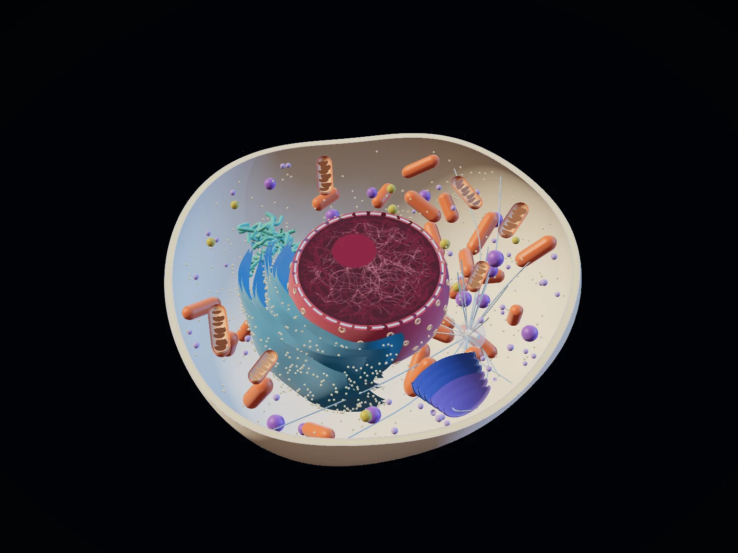
SpecimenAnimal cell
Field of view—
Typical size≈ 20 µm
Opening the 3D atlas…
Drag to orbit · Scroll to zoom · Select a structure
BIOLOGY / 001 · INTERACTIVE ATLAS
Life has an inner order.
Within a thin membrane, a world works together. Turn, uncover and observe: understand life through its structures.
THE COLLECTION / 001—008
A natural atlas you can turn
One original model collection, shared by the interactive views and the illustrated plates.
Model illustration, not a micrograph · Original modeling and production: OpenPith · 2026
FIELD NOTES
Science & image provenance
Original models created by OpenPith. Structural explanations draw on OpenStax Biology 2e, NCBI Bookshelf and Nobel Prize. Colors distinguish structures; they are not natural cell colors.
Light microscopy generally cannot resolve a lipid bilayer. Fluorescent labels reveal labeled structures only, and an electron micrograph of a section is not a complete 3D cell.
Cutaways, colors and structure counts serve teaching. Cells do not have these tidy viewing windows, and their interiors are dynamic.
From boundaries to cooperation
Enable JavaScript to rotate models. Cell structures and learning content remain readable below.
Cells are the fundamental units of life. Animal cells, plant cells and bacteria all have a plasma membrane, cytoplasm, ribosomes and genetic material, but organize their interiors differently.
Scale
From micrometers to nanometers. Begin with size, then understand why different tools reveal different structures.
One micrometer equals 1,000 nanometers. Cells, mitochondria and membrane thickness span orders of magnitude. Enlarging an image does not increase its resolution.
Cell sizes vary. These are typical orders of magnitude, not exact measurements of a species.
Overview
A cell is more than a bag of organelles: boundaries, information, energy and material flow form a system.
The membrane selectively exchanges material. Structures and molecules move through the cytoplasm. The nucleus, ribosomes and endomembrane system participate in gene expression and synthesis.
There is no single universal animal-cell shape. This model reduces organelle counts and widens the spaces between them.
Organelles
Mitochondrial cristae, chloroplast grana and ER membranes create working surfaces within a limited space.
Membranes compartmentalize reactions. Folds and stacks enlarge working surfaces, but membrane systems are not all freely connected.
Ribosomes and centrosomes are not membrane-bound. Textbooks differ in how broadly they use the term organelle.
Compare
They share fundamental components of life but organize them differently. The differences go beyond chloroplasts.
Animals and plants are eukaryotes. Bacteria lack a membrane-bound nucleus but have DNA, ribosomes and a plasma membrane. Leaf mesophyll cells typically have chloroplasts and a large vacuole.
The bacterium is schematic, not a complete E. coli envelope. Some bacteria lack a cell wall, and root cells usually lack chloroplasts.
Plasma membrane
Hydrophilic heads face water; hydrophobic tails gather inside. Proteins give this boundary selectivity.
Membranes are dynamic systems of lipids and proteins. Small nonpolar molecules cross lipid layers relatively easily; ions usually need channels or carriers.
Lipid counts and protein sizes are not to scale. The two leaflets differ in composition; carbohydrate chains generally face outward.
Transport
Passive transport follows gradients. Movement against an electrochemical gradient requires energy coupling. Bulk cargo moves in vesicles.
Channels and carriers help selected substances cross. Limited carrier numbers cause saturation. Each sodium–potassium pump cycle uses one ATP, typically sending 3 sodium ions out and 2 potassium ions in.
Ion movement depends on electrochemical gradients. The concentration controls model neutral solutes only. Secondary active transport uses an existing gradient rather than directly hydrolyzing ATP.
Osmosis
Net water movement changes cell volume. A plant wall limits swelling but cannot prevent the protoplast from shrinking when water leaves.
Under the assumptions used here, water moves toward the side with a higher effective solute concentration. Plant cells develop turgor in hypotonic conditions and may plasmolyze in hypertonic conditions.
Osmotic pressure, wall elasticity and membrane permeability jointly determine the outcome. The scaling animation is not a measured volume curve.
Secretion
From ribosomes to the ER and Golgi, a secreted protein is synthesized, processed, sorted and transported.
Proteins with suitable signals enter the ER, travel in vesicles to the Golgi, and are sorted to different destinations. Secretory vesicles eventually fuse with the plasma membrane.
Not every protein follows this route. The endomembrane system includes recycling and retrograde traffic; this view focuses on the forward secretory route.
Cytoskeleton
Microtubules and actin form dynamic networks. Motor proteins use energy to carry cargo such as vesicles along tracks.
Different motor proteins have different directional preferences. Microtubules, actin and their motors contribute to transport, shape changes and division.
Vesicles use only 7 microtubules, and pauses, detachments and track switches follow fixed rules. Kinesin steps 8 nm per ATP. Cytoplasmic streaming also involves actin–myosin mechanisms and should not all be attributed to microtubules.
Evidence
Distinguish observational evidence from explanatory models to learn from an image without being misled.
Stains, fluorescent labels and processing affect appearance. Check scale bars, labeled targets and imaging methods. Models should declare their selective simplifications.
Every plate here is rendered from an original 3D model, not acquired by microscopy. Fluorescence colocalization does not by itself prove direct molecular interaction.